| Cat # | Size | Price | Quantity | |
|---|---|---|---|---|
| 204903 | 25 tests | $75 | ||
| 204904 | 100 tests | $180 |
| Clone | RMP1-14 |
|---|---|
| Application | Flow Cytometry, IHC-F |
| Reactivity | Mouse |
| Format | iF647 |
| Target Name | CD279, Programmed Death-1, PD 1, PDCD1, PD-1 |
| Isotype | Rat IgG2a |
| Antibody Type | Monoclonal |
| Regulatory Status | RUO |
| Formulation | Phosphate-buffered solution, pH 7.2, containing 0.09% sodium azide and 0.2% (w/v) BSA |
| Protein Concentration | Supplied at a lot-specific concentration. |
| Storage&Handling | The antibody solution should be stored undiluted between 2°C and 8°C, and protected from prolonged exposure to light. Do not freeze. |
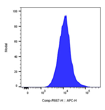
| Recommended Usage | For flow cytometric staining, it is recommended to use 5 uL of this reagent per 0.5-1.0 million cells in a 100 µL volume. Optimal reagent performance should be determined by titration for each specific application. iF647 has an excitation max at 656 nm and an emission max at 670 nm. |
| Excitation Laser | Red Laser (633 nm) |
| Isotype Controls | 303512 |
| Antibody Family | Mouse Antibodies |
| See All Formats | Clone RMP1-14 |
Mouse CD279, more commonly known as programmed cell death protein 1 (PD-1), is an inhibitory immune checkpoint receptor expressed primarily on activated T cells, as well as B cells and some myeloid populations. It plays a critical role in maintaining peripheral tolerance and preventing excessive immune activation by downregulating T cell responses during chronic antigen exposure, such as infection or inflammation. PD-1 is rapidly induced following T cell receptor engagement and acts as a key regulator of immune homeostasis.
Structurally, PD-1 is a type I transmembrane protein belonging to the immunoglobulin superfamily. It contains a single extracellular IgV-like domain responsible for ligand binding, a transmembrane region, and a cytoplasmic tail with immunoreceptor tyrosine-based inhibitory (ITIM) and switch (ITSM) motifs. Upon ligand engagement, these motifs recruit phosphatases such as SHP-2, which attenuate proximal T cell receptor signaling pathways.
The primary ligands of PD-1 are PD-L1 (CD274) and PD-L2 (CD273), which are expressed on antigen-presenting cells and various non-hematopoietic tissues. Interaction of PD-1 with its ligands suppresses T cell proliferation, cytokine production, and survival, promoting an exhausted T cell phenotype during chronic immune stimulation.
PD-1 signaling is implicated in chronic infections, cancer, and autoimmune diseases. In tumors, PD-1-mediated inhibition allows cancer cells to evade immune surveillance by suppressing anti-tumor T cell activity.
Therapeutically, blockade of the PD-1/PD-L1 axis using monoclonal antibodies has revolutionized cancer immunotherapy by restoring T cell function. Conversely, enhancing PD-1 signaling may be beneficial in treating autoimmune diseases and preventing transplant rejection, making it a versatile target in immune modulation.
iF647 Anti-mouse CD279 (PD-1) Antibody TDS
Have a product or application question? Consult our FAQs or contact us.